
Australian scientists achieve a milestone by visualizing the PINK1 protein, paving the way for innovative treatments for Parkinson’s disease.
Parkinson’s disease, a progressive neurodegenerative disorder, has long puzzled scientists, particularly concerning the role of the PINK1 protein. This protein is crucial for maintaining cellular health by identifying and marking damaged mitochondria for removal. Mutations in PINK1 disrupt this process, leading to the accumulation of dysfunctional mitochondria, which is especially detrimental to energy-demanding brain cells.
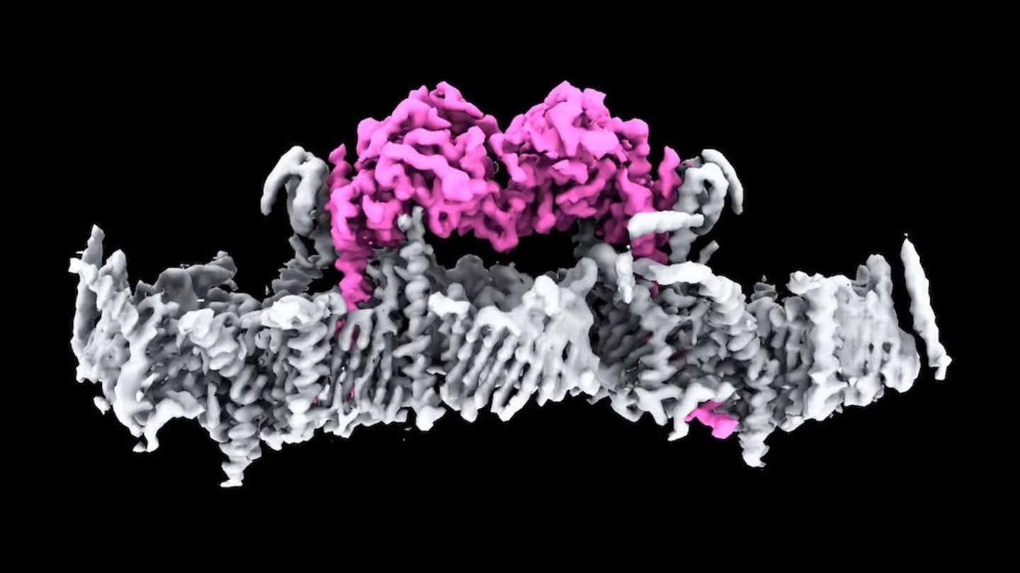
For over two decades, researchers have sought to understand the structure and function of PINK1. Now, a team at Australia’s Walter and Eliza Hall Institute (WEHI) has successfully imaged human PINK1 attached to mitochondria for the first time. Utilizing advanced cryo-electron microscopy, they unveiled how PINK1 binds to the mitochondrial surface and initiates the clearance of damaged components. This discovery also sheds light on how specific mutations associated with Parkinson’s impair PINK1’s function.
Professor David Komander, the study’s lead author, remarked on the significance of this achievement: “It is incredible to finally see PINK1 and understand how it binds to mitochondria. Our structure reveals many new ways to change PINK1, essentially switching it on, which will be life-changing for people with Parkinson’s.”
This breakthrough opens avenues for developing therapies aimed at reactivating or correcting PINK1 function, potentially slowing or halting the progression of Parkinson’s disease. While current treatments primarily address symptoms, targeting the underlying mechanisms offers hope for more effective interventions.
The research community is optimistic that these findings will accelerate the discovery of drugs capable of modulating PINK1 activity. Such advancements could transform the landscape of Parkinson’s treatment, offering renewed hope to those affected by this challenging condition.